

A 7-Week, Open-Label Study Evaluating the Efficacy and Safety of 415-nm/633-nm Phototherapy for Treating Mild-to-Moderate Acne in Adolescents and Adults
Ablon G.
Journal of Clinical and Aesthetic Dermatology. 2025;18(10):25–32
Randomized Trial of Three Phototherapy Methods for the Treatment of Acne Vulgaris in Chinese Patients
Li-Hong Liu, Xin Fan, Yu-Xi An, et al.
Photodermatol Photoimmunol Photomed 2014; 30: 246–253
A Study to Determine the Effect of Combination Blue (415 Nm) and Near-Infrared (830 Nm) Light-Emitting Diode (LED) Therapy for Moderate Acne Vulgaris
Sadick NS.
Journal of Cosmetic Laser Therapy. 2009;11(2):125–128
Handheld LED Array Device in the Treatment of Acne Vulgaris
Sadick NS.
Journal of Drugs in Dermatol. 2008;7(4):347–350.
Blue and Red Light Combination LED Phototherapy for Acne Vulgaris in Patients with Skin Phototype IV
Lee SY, You CE and Park MY.
Lasers in Surgery and Medicine. 2007; 39: 180-188
Light-Emitting Diode 415 nm in the Treatment of Inflammatory Acne
Tremblay JF, Sire DJ, Lowe NJ and Moy RL.
Journal of Cosmetic and Laser Therapy. 2006; 8: 31-33
Combination Blue (415 nm) and Red (633 nm) LED Phototherapy in the Treatment of Mild to Severe Acne Vulgaris
Goldberg DJ and Russell BA.
Journal of Cosmetic and Laser Therapy. 2006; 8: 71-75
An Open Study to Determine the Efficacy of Blue Light in the Treatment of Mild to Moderate Acne
Morton CA, Scholefield RD, Whitehurst C and Birch J.
Journal of Dermatological Treatment. 2005; 16: 219-223
Omnilux Contour Face Clinical Study Summary
Clinical Report
Omnilux Men Peer Reviewed Publication
Clinical Report
Visible Red Light Does Not Induce DNA Damage in Human Dermal Fibroblasts
Jennifer Y. Wang, Evan Austin, Jared Jagdeo
Clinical Report
Near-infrared Light Does Not Induce DNA Damage in Human Dermal Fibroblasts
Jennifer Y. Wang, Evan Austin, Jared Jagdeo
Clinical Report
The Use of Light-Emitting Diode Therapy in the Treatment of Photoaged Skin
Baez F and Reilly LR.
Journal of Cosmetic Dermatology. 2007; 6: 189-194
A prospective, Randomized, Placebocontrolled, Double-Blinded, and Split-Face Clinical Study on LED Phototherapy for Skin Rejuvenation: Clinical, Profilometric, Histologic, Ultrastructural, and Biochemical Evaluations and Comparison of Three Different Treatment Settings
Lee SY, et al.
Journal of Photochemistry and Photobiology B.2007; 88: 51-67
A Single-Blinded Randomized Controlled Study to Determine the Efficacy of Omnilux Revive Facial Treatment in Skin Rejuvenation
Bhat J, Birch J, Whitehurst C and Lanigan SW.
Lasers in Science. 2005; 20: 6-10
A Study to Determine the Efficacy of Combination LED Light Therapy (830 nm and 633 nm) in Facial Skin Rejuvenation
Russell BA, Kellett N and Reilly LR.
Journal of Cosmetic and Laser Therapy. 2005; 7: 196-200
Clinical trial of Non thermal 633nm Omnilux LED array for renewal of Photoaging: Clinical Surface Profilometric Results
Kim JW.
Journal of the Korean society for Laser Medicine and Surgery. 2005; 9: 69-76
A Near Infrared LED-Based Rehabilitation System: Initial Clinical Experience
Baxter GD, Bleakley C, Glasgow P and Calderhead RG.
Laser Therapy. 2005; 14.1: 29-36
Combined Infrared Laser and LED Therapy for Post Mastectomy Pain and Discomfort: A Case Report
Trelles MA and Calderhead RG.
Phototherapy With Light Emitting Diodes: Treating a Broad Range of Medical and Aesthetic Conditions in Dermatology
Ablon G.
Journal of Clinical Aesthetic Dermatol. 2018;11(2):21–27.
The Photobiological Basics Behind Light-Emitting Diode (LED) Phototherapy
Calderhead RG.
Laser Therapy. 2007; 16.2: 97-108
Light-Emitting Diode Phototherapy at 630 +/- 3 nm Increases Local Levels of Skin-Homing T-Cells in Human Subjects
Takezaki S, Omi T, Sato S and Kawana S.
Journal of Nippon Medical School. 2006; 73:75-81
Ultrastructural Observations Of Human Skin Following Irradiation With Visible Red Light-Emitting Diodes (LEDs): A Preliminary In Vivo Report
Takezaki S, Omi T, Sato S and Kawana S.
Laser Therapy. 2005; 14.4: 153-160
Laser Resurfacing Today – Not All Photoscience is Photothermal
Calderhead RG.
Journal of Cosmetic Dermatology. 2004; 2: 242-243 (Letter to the Editor)
One Mechanism Behind LED Phototherapy for Wound Healing and Skin Rejuvenation: Key Role of the Mast Cell
Calderhead RG, Kubota J, Trelles MA and Ohshiro T.
Laser Therapy. 2008; 17.3: 141-148
Low Level Laser Therapy and Phototherapy Assisted Hydrogel Dressing in Burn Wound Healing: Light Guided Epithelial Stem Cell Biomodulation
Kim JW and Lee JO.
In: Innovations in Plastic and Aesthetic Surgery. Springer; 2008. p. 36-41
Combined Visible Light and Infrared Light-Emitting Diode (LED) Therapy Enhances Wound Healing After Laser Ablative Resurfacing of Photodamaged Facial Skin
Trelles MA, Allones I and Mayo E.
Medical Laser Application. 2006; 21: 165-175
Red Light-Emitting Diode (LED) Therapy Accelerates Wound Healing Post-Blepharoplasty and Periocular Laser Ablative Resurfacing
Trelles MA and Allones I.
Journal of Cosmetic and Laser Therapy. 2006; 8: 39-42
Red Light-Emitting Diode (LED) Therapy-Assisted Healing Improves Results of Er:Yag Laser Ablation of Plantar Verrucae
Trelles MA and Calderhead RG.
Laser Therapy. 2005; 14.4: 179-183
The improvement of Hypertrophic Scar and Keloidal Scar by Combining Drilling Tiny Pinholes with Carbon Dioxide Laser and 830nm Omnilux PDT LED
Kim JW, Lee JO and Calderhead RG.
Journal of the Korean society for Laser Medicine and Surgery. 2005; 9: 1-6
Synergic Effect of Ablating Erbium Yag Laser Skin Resurfacing Combined with Non-Ablating Quasilaser Light 415nm, 633nm, 830nm LED Array in Asian Patients
Kim JW, Lee JO, Calderhead RG, Rhee CG and Ahn WS.
Journal of the Korean Society for Laser Medicine and Surgery. 2005; 9: 15-23
* Please Note: Wound Healing & Photodynamic Therapy are not FDA-cleared indications.[1]
Pilot Study to Determine the Efficacy of ALAPDT Photorejuvenation for the Treatment of Facial Aging
Lowe NJ and Lowe P1.
Journal of Cosmetic and Laser Therapy. 2005; 7: 159-162
* Please Note: Wound Healing & Photodynamic Therapy are not FDA-cleared indications.[1]
Long-term Follow-Up of Photodynamic Therapy With a Self-Adhesive 5-aminolaevulinic Acid Patch: 12 Months Data
Szeimies RM, Stockfleth E, Popp G, et al.
British Journal of Dermatol. 2010;162(2):410–414
Optimization of Photodynamic Therapy with a Novel Self-Adhesive 5-Aminolaevulinic Acid Patch
Hauschild A, Stockfleth E, Popp G, et al.
British Journal of Dermatol. 2009;160(5):1066–1074
Efficacy of ALA-PDTvs. Blue Light in the Treatment of Acne
Akaraphanth, R, et al.
Photodermatology Photoimmunology & Photomedicine 2007; 23: 186–190.
In Vitro and In Vivo Comparison of Two Different Light Sources for Topical Photodynamic Therapy
Babilas P, et al.
British Journal of Dermatology. 2006; 154: 712-718.
Comparison of Paterson PDT and LED Light Outputs
Hutson S.
Internal document of Photo Therapeutics Ltd. DES/Tech/0506. May 28, 2002
Photodynamic Therapy for Large or Multiple Patches of Bowen’s Disease and Basal Cell Carcinoma
Morton CA, Whitehurst C, McColl JH, Moore JV and MacKie RM;
Archives of Dermatology. 2001; 137: 319-324
Treating Basal Cell Carcinoma: Has Photodynamic Therapy Come of Age?
Morton CA.
British Journal of Dermatology. 2001; 145: 1-2 (editorial comment)
Routine Double Treatments of Superficial Basal Cell Carcinomas Using Aminolaevulinic Acid Based Photodynamic Therapy
Haller JC, Cairnduff F, Slack G, Schofield J, Whitehurst C, Tunstall R, Brown SB and Roberts DJH.
British Journal of Dermatology. 2000; 143: 1270-1274
Comparison of Red and Green Light in the Treatment of Bowen’s Disease by Photodynamic Therapy
Morton CA, Whitehurst C, Moore JV and MacKie RM.
British Journal of Dermatology, 2000, 143, 767-772.
Photodynamic Therapy with Meso-Tetra (hydroxyphenyl) Chlorin in the Topical Treatment of Bowen’s Disease and Basal Cell Carcinoma
Gupta G, Morton CA, Whitehurst C, Moore JV and Mackie RM.
British Journal of Dermatology. 1999; 141: 385-386
Treatment of Penile Lichen Planus with Photodynamic Therapy
Kirby B, Whitehurst C, Moore JV and Yates VM.
British Journal of Dermatology.1999; 141: 765-766 (correspondence).
Photodynamic Therapy for Basal Cell Carcinoma – Effect of Tumor Thickness and Duration of Photosensitizer Application on Response
Morton CA, Whitehurst C, McColl JH, Moore JV and Mackie RM.
Archives of Dermatology. 1998; 134: 248-249
Comments on: Problems Associated with the Use of Broad-Band Illumination Sources for Photodynamic Therapy
Roberts F, Whitehurst C and Moore JV.
Physics in Medicine and Biology. 1996, 41, 1518-1521
Comparison of Photodynamic Therapy with Cryotherapy in the Treatment of Bowen’s Disease
Morton CA, Whitehurst C, Moseley H, McColl JH, Moore JV and Mackie RM.
British Journal of Dermatology. 1996; 135: 766-771
Photodynamic Therapy of Primary Skin Cancer: A Review
Roberts DJH and Cairnduff F.
British Journal of Plastic Surgery. 1995; 48: 360-370
Development of an Alternative Light Source to Lasers for Photodynamic Therapy: 3. Clinical Evaluations in the Treatment of Pre-Malignant Nonmelanoma Skin Cancer
Morton CA, Whitehurst C, Moseley H, Moore JV and MacKie RM.
Lasers in Medical Science. 1995; 10: 165-171
Development of an Alternative Light Source to Lasers for Photodynamic Therapy: 2. Comparative In Vivo Tumor Response Characteristics
Whitehurst C, Humphries JD and Moore JV.
Lasers in Medical Science. 1995; 10: 121-126
Development of an Alternative Light Source to Lasers for Photodynamic Therapy: 1. Comparative In Vitro Dose Response Characteristics
Whitehurst C, Byrne K and Moore JV.
Lasers in Medical Science. 1993; 8: 259-267
* Please Note: Wound Healing & Photodynamic Therapy are not FDA-cleared indications.[1]

Blog
View our blog covering all things light therapy, by the leaders in light therapy.LED light therapy

LED phototherapy
LED (Light Emitting Diode) devices are well established in clinical anti-aging and skin-care therapy, as well as in the treatment of a wide variety of skin conditions. Omnilux™ Professional LED devices are the gold-standard medical technology used by dermatologists and aestheticians world over because of their ability to promote skin recovery and healing.
Omnilux products combine clinically proven wavelengths of light with optimized energy levels in a process called LED phototherapy.
Visit Website
Here’s how it works
When you use an Omnilux product, the LEDs emit light into your skin.
Light photons of specific wavelengths are absorbed by cell components, particularly the mitochondria, triggering a cascade of biochemical reactions as explained in this video.

SEE HOW IT WORKS +

As we age, collagen and elastin naturally decline, leading to volume loss. Skin may appear more wrinkled, loose, thin, crepey, and unevenly textured. Years of environmental exposure can accelerate visible signs of aging on the face, neck, chest, and hands.
Red and near-infrared wavelengths work on a cellular level to stimulate fibroblast activity, support collagen and elastin production, improve elasticity, and reduce the appearance of fine lines and wrinkles.
The red wavelength (633nm) supports cellular repair and circulation, promoting a more vibrant complexion. The near-infrared wavelength (830nm) penetrates deeper layers of the skin to support collagen and elastin production, resulting in plumper, firmer, more youthful looking skin.
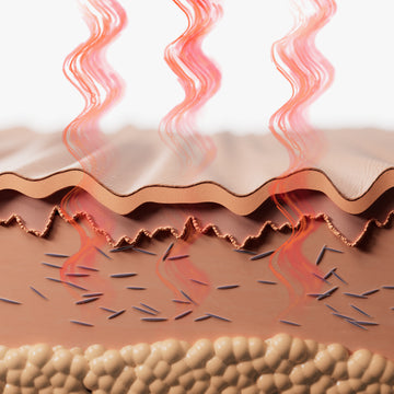
Visible red and near infrared light
is absorbed into the skin and
dermis.
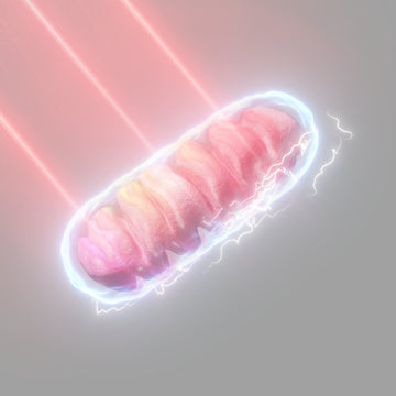
Mitochondria (the cell's "battery") absorb the light and convert it to energy for cellular activity.
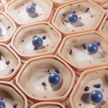
Increased cellular activity produces a cascade of reactions called "secondary reactions".
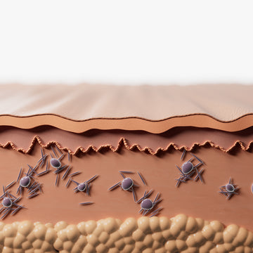
Secondary reactions include the increased production of collagen (called collagenisis) and elastin.
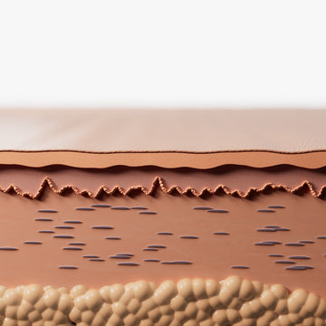
New collagen forms within the dermis, resulting in plumper, smoother and more radiant skin.
The results are visibly smoother, firmer skin with a reduction in fine lines and wrinkles and a more even skin tone.

Before Omnilux Contour:
Uneven texture and visible signs of aging.

After Omnilux Contour:
Skin appears smoother, plumper, and brighter.

SEE HOW IT WORKS +

Acne begins when the sebaceous glands produce excess oil. This oil combines with dead skin cells, clogging pores and contributing to breakouts. Certain skin bacteria, including C. acnes, produce natural chemicals called porphyrins, which are sensitive to the blue and red wavelengths used by Omnilux Clear.
The blue light (415nm) targets and neutralizes acne-causing bacteria on the skin’s surface. Combined with red light (633nm), Omnilux Clear helps reduce active acne breakouts by supporting the skin’s natural recovery process.
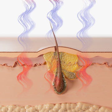
Visible blue & red light is absorbed into the skin and dermis.
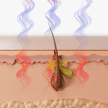
Blue light penetrates the superficial levels of the skin, eliminating acne causing bacteria called porphyrins (P.acne).
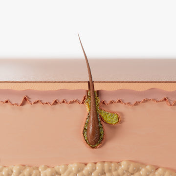
Deeper penetrating red light stimulates an anti-inflammatory response, reducing redness and stimulating a healing response.
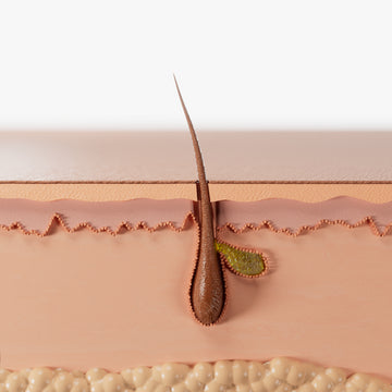
A completely non-invasive and natural treatment - Omnilux Clear uses the skin’s natural responses to treat acne, leaving clear and brilliant skin.
The results are visibly smoother, clearer skin with a more even tone and a reduction in acne.

Before Omnilux Clear:
Pronounced acne and visible scarring.

After Omnilux Clear:
Noticeable reduction in acne, smoother skin texture, and a more vibrant complexion.

Noninvasive and gentle on the skin
LED therapy is a completely natural treatment and does not contain any harmful UV wavelengths; it does not create heat or thermally damage the skin.

Clinically proven wavelengths for optimal results
Each wavelength, or color of light, stimulates a specific skin response:
- Red (633nm): Supports cellular repair and circulation, promoting visibly healthier, more radiant skin.
- Near‑infrared (830nm): Targets deeper fibroblast cells to stimulate collagen and elastin production for plumper, firmer, more youthful looking skin.
- Near‑infrared (1072nm): Penetrates deeper into men’s thicker skin, triggering a robust cellular response that supports anti-aging and skin rejuvenation.
- Blue (415nm): Targets and neutralizes acne-causing bacteria on the skin’s surface to help reduce active breakouts.